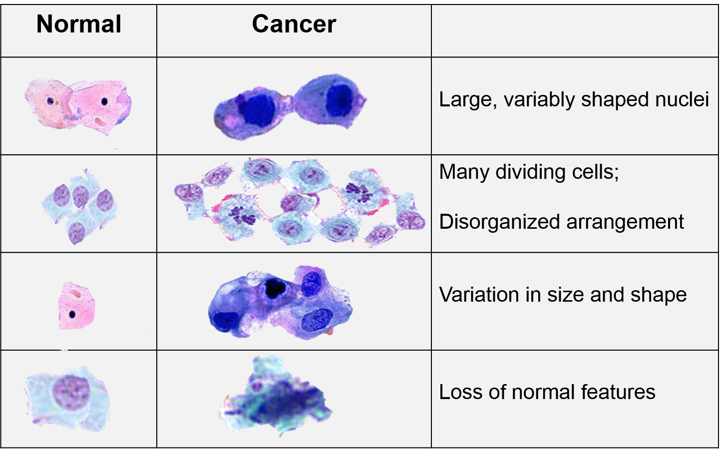
Used to Describe the Histological Characterization of Tumor Cells
Rare cases of tumors with predominant osteoclast-type giant cell histology have been reported see below and tumors with focal osteoclast-like giant cells should not be misclassified as these. Hypoxia in tumors signifies resistance to therapy.

H E Staining Of Tumor Cells Abbreviation H E Hematoxylin And Eosin Download Scientific Diagram
Essentially cancer is a disease of mitosis.

. Transitional cell describes the morphology of the urothelium but transitional cells can also be found in other anatomic locations so. Cell-cycle quiescence might be a common mechanism underlying the long-term maintenance of stem-cell function in normal and neoplastic stem cells and our previous study demonstrated that quiescence induced by hypoxia-inducible factor. They are frequently used in the detection and diagnosis of skin cancer.
Despite a wealth of tumor histology data including anti-pimonidazole staining no current methods use these data to induce a quantitative characterization of chronic tumor hypoxia in time and space. Characterization of the niches for stem-like tumor cells is important to understand and control the behavior of glioblastomas. Essentially breast cancer histology evaluation is the microscopic analysis of the chemical and cellular properties of the cells of a suspicious breast tumor.
Histological characterization of tumor vessels is also important because angiogenesis is one of the key processes in tumor growth and progression8 Normalizing abnormal growth of. FOXP3 regulatory T-cells were observed in only 466 6 tumors all of which were astrocytomas. Molecular and histological profiling of lung cancer is important to identify molecular and.
Biomarkers on cancer cells is one of the main indicators of a neoplastic process which may be facilitated by using antibodies andor aptamers. Lung cancer is the most common and deadly cancer accounting for about 176 million deaths globally. Histology refers to the study of the individual parts and structures which make up a cell and the relationship between structure and function.
While histomorphologic assessment provides important prognostic information genomic analysis of tumors can provide important. As such it occurs when normal cells are transformed into cancerous cells and proliferate uncontrollably. Visual comparison highlights the trained networks ability to distinguish histological.
Tumor Node Metastasis system is. Cancer cells therefore are normal cells whose genes several genes have been damagedmutated which in turn cause the cell as a whole to respond differently to signals that control the lifespan. Histology and histopathology of biopsy samples are important in the diagnosis of skin conditions.
The presence of variant histology may be associated with more advanced stage at presentation and a poorer response to systemic therapy. Acceleration of the cell cycle. So breast cancer histology is essential to determine the most effective approaches to.
The language used to describe conventional bladder cancer has evolved from the more traditional term transitional cell carcinoma to the now more commonly used term urothelial carcinoma 23. Secretion of lytic factors etc. Histological and immunohistochemical characterization of feline renal cell carcinoma.
The malignant cell is characterized by. Researchers imaging data along with image-processing algorithms in order to develop a set of candidate image features that can be used to develop a quantitative description of xenografted colorectal chronic tumor hypoxia. The purposes of this study are to describe the histological features.
In an attempt to incorporate proteomics into a biological understanding of pediatric brain tumors we undertook the first large-scale comprehensive proteogenomics analysis inclusive of the genomics transcriptomics and global and phosphoproteomics of a large cohort of 218 tumor samples representing 7 distinct histological diagnoses including low. A case series. White space misclassified as other cellular classes may be due to the presence of lumen Fig.
What are histology stains. CD8 T-cells infiltrated 476572 tumors irrespective of their histology or IDH1 mutation status. Immunohistochemistry and regular histology were used to describe features such as tumor cell infiltration necrosis area nuclear pleomorphism cellularity mitotic characteristics leukocytic infiltration proliferation and inflammation.
Histological Characterization of Some Feline Mammary Gland Tumors with Whole Slide Images Scan as a Trial of Remote Diagnosis. Ovarian cancer is the most common cause of elevated CA 125 but cancers of the uterus cervix pancreas liver colon breast lung and digestive tract can also raise CA. Transitional cell describes the morphology of the urothelium but transitional cells can also be found in other anatomic locations so.
They were seen more often in wild-type than in mutant IDH tumors P 0046 MannWhitney U-test. These features were used to develop a spatiotemporal logical expression as well as a way to formulate a linear regression function that uses all of the image. Cancer and Cancer Cells.
Changes in the cellular surface. Tumor cell class which was most likely a biological result of short straight fibers amongst tumor cells. Colorectal cancer is the most common cancer where this tumour marker is used but many other epithelial cancers can also raise levels.
The pathologist will also confirm the size of the breast tumor where necessary for breast cancer staging purposes. Up to 25 of tumors are of pure variant or mixed urothelial and variant histology. In an attempt to incorporate proteomics into a biological understanding of pediatric brain tumors we undertook the first large-scale comprehensive proteogenomics analysis inclusive of the genomics transcriptomics and global and phosphoproteomics of a large cohort of 218 tumor samples representing 7 distinct histological diagnoses including low.
Compared to primary. The language used to describe conventional bladder cancer has evolved from the more traditional term transitional cell carcinoma to the now more commonly used term urothelial carcinoma 23.

Biological Characteristics Of Malignant Cells A Histology Of Human Download Scientific Diagram

Characteristics Of Cancer Cells

Biological Characteristics Of Malignant Cells A Histology Of Human Download Scientific Diagram
Comments
Post a Comment